Douglas H. Ubelaker1
1. Department of Anthropology Smithsonian Institution. United States. E.mail: ubelaked@si.edu
Tipologia: Articulo de reflexión
Fecha de recepción: 21/01/2014
Fecha de aceptación: 18/06/2014
Como citar éste artículo: Ubelaker, D. H. (2014). Contributions of Pathological Alterations to Forensic Anthropology Interpretation. Jangwa Pana.13, 140 - 151
Pathology plays a key role in various aspects of interpretation within forensic anthropology. These contributions include observations of the effects of pathological conditions and using them to facilitate identification efforts. In the context of the broader definition of pathology as "something abnormal" it forms the foundation of much of the logic, methodology and practice of forensic anthropology. In fact, only unusual conditions not shared by many others can be utilized for positive identification. Recovery efforts and evaluation of the evidence of foul play, as well as the estimation sex, age at death, ancestry, living stature, time since death and other important components depend upon recognition of normal and abnormal patterns. The multifaceted contributions of pathology to the practice of forensic anthropology are firmly documented in casework applications.
Keywords: Pathology, identification, forensic anthropology
La patología juega un rol principal en varios aspectos relacionados con la interpretación dentro de la antropología forense. Se presentan aquí diversas observaciones sobre los efectos de las condiciones patológicas y las formas cómo éstas pueden ser usadas para facilitar el proceso de identificación. Es justamente en el contexto de la definición más amplia de patología, como "algo anormal", que se forma en gran medida la base de la lógica, de la metodología y de la práctica de la antropología forense. De hecho, las condiciones que no son frecuentes o que no se comparten con muchas otras personas son las que pueden ser utilizadas para llegar a la identificación positiva. Los esfuerzos durante el rescate y la evaluación de la evidencia de actividad criminal (foul play), así como la estimación del sexo, la edad al momento de la muerte, la filiación poblacional, la estatura en vida, el tiempo transcurrido desde el momento de la muerte y otros componentes importantes, dependen del reconocimiento de los patrones normales y los anormales. Las múltiples contribuciones de la patología a la práctica de la antropología forense son documentadas aquí a través de casos aplicados.
Palabras clave: Patología, identificación, antropología forense
Forensic anthropology represents the application of knowledge and methodology in anthropology to the resolution of medicolegal issues. Such issues can involve living individuals and those recently deceased but usually relate to largely skeletonized human remains. These contributions can include recovery of human remains, determination if recovered materials are human or non-human, estimation of sex, age at death, ancestry and living stature and evaluation of time since death. However, the central goals are concentrated on individual identification and evaluation of evidence of possible foul play. Although many lines of evidence and observations can be utilized, pathological alterations frequently provide key data for interpretation. This essay explores the multifaceted role of pathology in forensic anthropology investigation.
The word pathology usually is thought of as relating to the study of the disease process and its effects. In fact, the first definition of pathology in Webster's dictionary (Webster's, 1979) reads "the study of the essential nature of diseases and esp. of the structure and functional changes produced by them." This definition relates strongly to the general field of paleopathology and many applications within forensic anthropology. In the study of the human skeleton, abnormal alterations may be discovered. Interpretations of these alterations may raise the question "What disease process produced these alterations?" With ancient skeletons recovered from archeological contexts, the question is important to examine the history, evolution and impact of disease and to explore general past patterns of morbidity and mortality (Waldron, 2009). In the forensic context, an accurate answer to the question may lead to family memories of the disease experience, hospital records and identification. However, answers may remain elusive since many different diseases produce similar effects on the skeleton and single diseases may have multiple, distinctive effects (Aufderheide and Rodriguez-Martin, 1998).
The second definition of "pathology" in Webster's dictionary (Webster's, 1979) reads simply "something abnormal." While such an abnormal condition may relate to disease it also may simply consist of a "deviation from property or from an assumed normal state..." This condition of "abnormality" rests at the very intellectual heart of forensic anthropology, especially as it relates to interpretations of personal identification and evidence of foul play. Positive identification depends upon the finding of unusual unique features on the recovered remains that can be demonstrated to have been present in a particular individual during life (Christensen and Anderson, 2013). Similarly, strong evidence for foul play must relate to abnormal alterations found on remains that were produced from events at or about the time of death (Berryman et al., 2013a,b). The concept of abnormality inherent within definitions of pathology also frames the core of anthropological interpretation, especially in relation to identification and foul play issues.
Contributions of forensic anthropologists to the determination of cause and manner of death usually involve observations of perimortem trauma (Loe, 2009). The term "perimortem" is employed in reference to the timing of the event that produced the observed alterations. Perimortem means at or about the time of death. Such a vague definition reflects the fact that from skeletal evidence alone, anthropologists are not able to define the exact timing relationship between death and the trauma event. To some extent, perimortem status represents a default category; the alterations lack evidence of antemortem remodeling/ new bone formation and present no diagnostic indications of postmortem status. Common postmortem indicators include coloration contrasts between the broken and unbroken surfaces, tool marks produced during recovery procedures, alterations diagnostic for chewing by rodents and other mammals, and alterations produced by plant root growth or sun exposure (Haglund and Sorg, 1997; Nawrocki, 2009).
In addition, evidence for perimortem timing may involve characteristics indicating the bone was in a fresh state when the alterations were sustained. Such evidence relates to a plastic response of fresh bone, such as the tendency to bend or curl. In research comparing breakage patterns in fresh bone vs. dry bone, Moraitis and Spiliopoulou (2006) found that the fresh bone pattern involved obtuse or acute fracture angles. They also noted that the fresh bone fracture edges were smoother, sharper and more beveled than those of dry bone. The term "trauma" in forensic anthropology context is closely linked with the perimortem definition provided above. Alterations considered to be traumatic do not include the bone breakage and weathering associated with postmortem factors. The types of perimortem trauma commonly observed in forensic anthropology casework involve blunt force trauma, sharp force trauma, gunshot injury, blast trauma and thermal effects (Loe, 2009). Although these categories can overlap, each presents particular diagnostic features. Interpretation reflects understanding of the nature of the types of force applied as well as the biomechanical properties of bone (Ubelaker, 1991). All forms of perimortem trauma present abnormal alterations and evidence suggesting they do not reflect normal human anatomical variation and were not produced long after death. Interpretation of perimortem trauma then calls for recognition of the patterns of alterations associated with the categories listed above, in conjunction with other evidence (e.g. context and recovery of projectiles). The following presents examples of the primary forms of perimortem trauma observed in forensic anthropology practice.
The term blunt force trauma refers to perimortem injuries that are general in nature and cannot be linked to more specific patterns or types of injury. Blunt force trauma can result from injury associated with blows with objects, vehicle crashes and falls from considerable heights. Their proper interpretation calls for the assessment of perimortem status as discussed above and also recognition of patterns and employment of biomechanical principles.
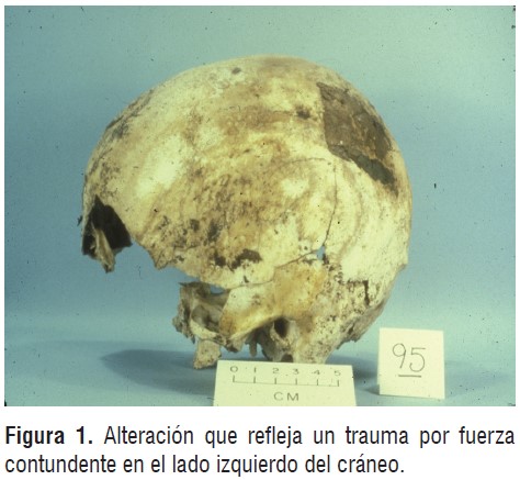
La Figure 1 presents an alteration on the left side of a cranium reflecting blunt force trauma. The affected area is slightly pushed inward with fracture borders. While the area is suggestive of perimortem blunt force trauma, the alterations do not reveal sufficient detail for a more precise diagnosis.
In some cases, the injuries sustained in blunt force trauma are sufficiently patterned that they can be linked to a particular type of object. Such "patterned trauma" may suggest class characteristics or general information regarding the nature and form of the materials involved. The extent to which such patterns can be detected depends not only on the instrument employed but also on the areas of the skeleton impacted.
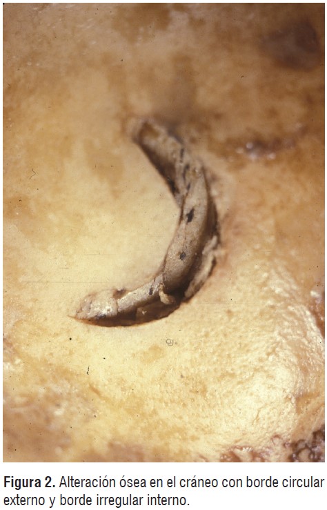
In 1990, a dispute in rural Washington State led a man to strike another with the working end of a tire iron (Ubelaker and Scammell, 1992).
Although an attempt was made to dispose of the body by putting it into a nearby river, eventually it was recovered and made available for analysis. Careful examination revealed alterations to the cranium suggestive of impact with a hard object. In particular, one of the alterations (Figure 2) revealed a circular outer border and an irregular inner border. The measurements and pattern of this alteration closely matched those of one end of a tire iron that had been recovered from the scene. My report and subsequent court testimony noted the similarities and indicated that the cranial alterations were produced either by that instrument or another one with the same features.
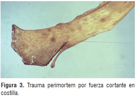
Sharp force trauma represents another form of injury in which the features of the impacted tissue suggest that an edged instrument was involved.
Such alterations tend to be linear with well-defined margins (Ubelaker, 1999). Since many different instruments can inflict sharp force trauma, the form of the alterations can vary extensively. Some can be very long, narrow and superficial while others can penetrate deeply into the bone or even separate it into segments. While injury variation can be profound, all present evidence that an edged instrument was involved.
La Figure 3 presents an example of perimortem sharp force trauma in a rib. Note the linear nature of the alteration, the clearly cut margins and the bending of the partially detached segment. While the nature of alteration suggests sharp force trauma, the outward bending of the segment indicates perimortem status. The outward bending was possible because the bone was in a fresh state when the injury occurred.
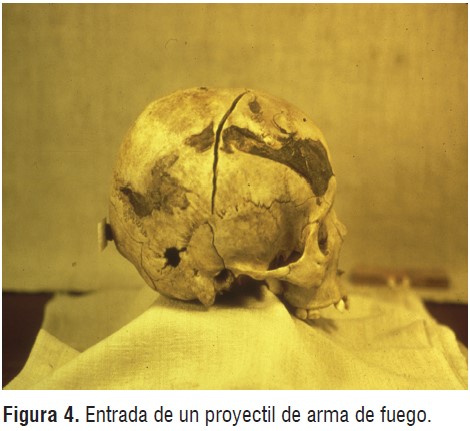
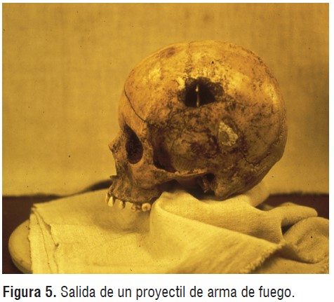
Gunshot injury may produce diagnostic skeletal alterations, depending upon the bone impacted. Projectile impact on dense cortical bone, such as that of the adult cranial vault may result in patterns of beveling that indicate direction. Entry gunshot injury may produce a larger perforation on the endocranial (internal) surface than on the corresponding ectocranial (external) surface (Figure 4). This pattern is reversed at the exit site (Figure 5). If the projectile strikes cancellous bone, the pattern of alteration may be more irregular. Direction may be evaluated through consideration of the beveling patterns discussed above, along with any evidence of fragment displacement (Ubelaker, 1996). Of course, evidence may also involve perforations in associated clothing or recovery of the projectiles themselves.
Blast trauma and thermal alterations both produce fragmentation. Blast trauma is frequently associated with thermal alterations thus these types of trauma are closely related. Thermal alterations usually present coloration and/or evidence of bone reduction that are diagnostic (Ubelaker, 2009). Determination of perimortem vs. post mortem status can be problematic in such cases since many taphonomic factors can also lead to fragmentation. Bones fragmented postmortem can also be exposed subsequently to thermal alteration.
Detection and interpretation of pathological alterations on the human skeleton must consider and rule-out those produced postmortem. Postmortem alterations can best be detected when they produce marked coloration contrasts between the broken and unbroken surfaces. Such contrast occurs when the break is relatively recent, such as those sustained during recovery. A fresh postmortem break of a bone that has been exposed to the elements for many years usually will reveal the natural bone color and texture in the broken surface. These indicators may contrast dramatically with the stained and weathered unbroken bone surfaces.
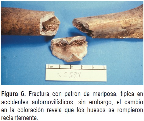
In some cases, the pattern of skeletal injury may be suggestive of perimortem trauma, but the contrasts in coloration can clarify postmortem status. In 1993, a construction worker in Georgia noted during site preparation that apparent skeletal remains had been uncovered. Analysis confirmed the human status of the remains and noted a series of fractures of the long bones suggestive of perimortem trauma. In particular, the fractures revealed a "butterfly" pattern in which roughly triangular areas of bone had been separated from the other areas of the diaphyses through fracture. This pattern of fracture has been observed in perimortem trauma involving pedestrians being struck by automobiles. In such cases the legs are usually involved. The bone impact site involves compression stresses that lead to complex fractures. Tension stresses are produced on the opposite side of the bone (non-impact surface) resulting in simple fractures of bone separation.
While the pattern of fractures in the Georgia case presented those suggestive of the perimortem trauma described above, the broken bone surfaces clearly revealed coloration contrasts. While the unbroken outer bone surfaces were uniformly stained due to postmortem exposure, the broken surfaces were unstained and revealed the natural color of bone (Figure 6). Apparently, the bones had been broken recently by the trampling effect of the weight of the heavy power equipment employed at the construction site (Ubelaker and Adams, 1995). This recent trampling effect had mimicked a well-known perimortem trauma condition. The case reveals the challenges of forensic interpretation and the value of careful examination for taphonomic indicators.
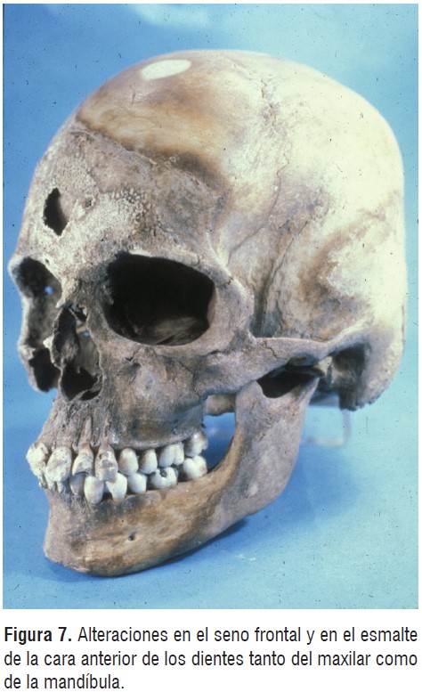
Another example is provided by Ubelaker and Sperber (1988) in their report on a forensic case from Omaha, Nebraska. A 19 year old female from Omaha had been reported missing in 1975, last seen attending a local gathering of the notorious Hell's Angels motorcycle organization. In 1984, a human skeleton was discovered within an unused cistern at the local airport and subsequently identified as the missing woman. Analysis confirmed the identity through dental records and noted a pattern of unusual perimortem alterations on the bones of the anterior skull suggestive of the application of a corrosive agent. These alterations were extreme exposing the frontal sinus and producing considerable alteration of the dental enamel in both the maxillary and mandibular anterior dentition(Figure 7).
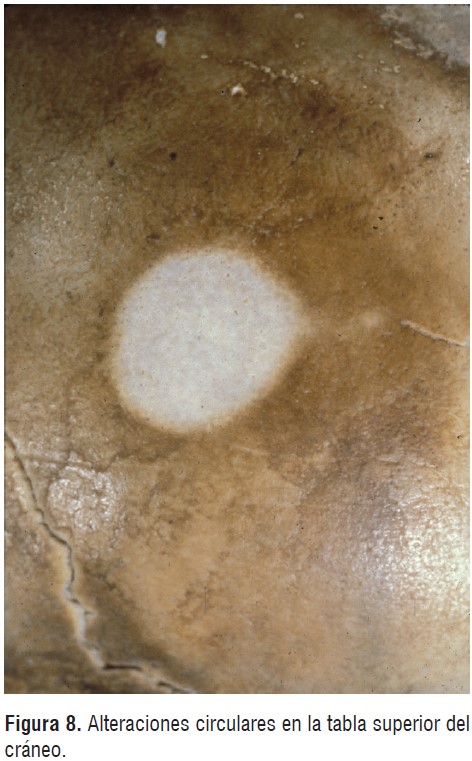
Analysis of the Nebraska case also noted circular alterations on the superior aspect of the cranium (Figure 8). While these alterations were similar in coloration to the corrosive destructive areas in the facial area, they did not present evidence of bone destruction. Final interpretation suggested these circular alterations had been formed postmortem through long term restricted sun exposure focused on the cranium through circular perforations in the cistern cover. Although the circular alterations were similar in coloration to those produced on the facial area, they were clearly postmortem and unrelated.
Many taphonomic alterations represent wellknown postmortem factors and can be easily recognized. Chew marks by rodents and other mammals can produce diagnostic bone alterations. Plant root growth can produce bone fracture and leave characteristic markings on bone surfaces. Sun exposure, fungal and algae growth, staining by decomposing leaves, soil contact and interaction with associated metal artifacts all represent well-known taphonomic factors (Nawrocki, 2009). Many of the alterations produced by these agents can mimic perimortem conditions, thus calling for experienced interpretation.
An important skill that forensic anthropologists bring to casework is knowledge of normal anatomical variation. Such information is vitally needed to recognize abnormal attributes and to avoid confusing normal features with them. Normal variation consists of developmental anatomical details that are known to represent natural human variation. Within such variation, anatomical features can be associated with general occurrence in different populations.
When features are found that fall outside of normal variation or are known to be rare, they acquire special value in forensic analysis. Such unusual features may provide key information contributing to identification. Some aspects of anatomy are known to exhibit great variation in morphological expression; so great that each individual example offers potential information for identification. The frontal sinus structure represents a case in point. The bony structure of the frontal sinus is known to exhibit such variation that even identical twins will show some differences (Ubelaker, 1984). As such this area of anatomy has proven its worth in personal positive identification efforts. Other areas of skeletal anatomy offer similar potential if the relevant remains are recovered and the corresponding antemortem radiographs become available.
The issue of uniqueness is paramount within forensic science. Within forensic anthropology, this issue represents a dominant feature of personal identification. The identification process represents a two-step process. In the first step, anatomical features must be found that are shared between the recovered remains and the documented antemortem information of a missing person. These features usually consist of anatomical features such as the frontal sinus discussed above. The antemortem information usually involves radiographs taken of the individual before death.
The second critical step involves assessing the uniqueness of the features shared. To support positive identification, the features must be unique or known to be shared with so few other individuals that the likelihood of the remains relating to another person is extremely remote. Usually such evaluation rests upon a suite of features that collectively are judged to be unique.
For example, the technique of photographic superimposition involves judging the relationship between anatomical features of a recovered skull with antemortem facial photographs of a missing person (Ubelaker, 1994). The modern equipment available today to facilitate this comparison involves computer imaging and represents a sophisticated and impressive process. In my experience, this technique is very useful for exclusion. When similarities are found and the missing person cannot be excluded, usually the report indicates that the remains might represent (cannot exclude) the missing person but positive identification cannot be achieved using this technique. While positive identification is theoretically possible, I have never applied this technique in a forensic case producing positive identification. Basically, it cannot be demonstrated that the shared features, as visually impressive as they might be, actually are unique.
In contrast, positive identification can be achieved when unique features are visible on antemortem radiographs. In such a case a cranial fragment was recovered in a New England homicide investigation and thought to represent a particular missing person. Careful detective work discovered that shortly before she died, the young woman had entered a local health clinic seeking relief from a severe headache. Officials at the clinic had taken cranial radiographs that were available for comparison. Radiographs taken of the recovered remains revealed shared morphology in the frontal sinus, sella turcica area of the sphenoid and other features that were in my opinion collectively unique enabling positive identification (Ubelaker, 1984).
In another case, positive identification was achieved through comparison of chest radiographs with recovered remains. In 1984, the largely skeletonized remains of an adult male were recovered recovered on an American Indian Reservation in South Dakota. Following anthropological analysis of the skeleton, authorities suggested the remains might relate to a known missing person. Although the dentition presented numerous restorations, efforts to locate antemortem dental records failed. The search did discover antemortem chest radiographs that were available for comparison. Comparison of the recovered remains with skeletal details visible in the radiographs resulted in positive identification (Ubelaker, 1990).
Another example reveals how unique skeletal features revealed in radiographs can be used to positively identify living individuals. This case involves Workers' Compensation Fraud on the part of members of a terrorist organization in the United States. Investigation revealed that in order to raise financial support for their organization, members with pre-existing ailments would accept employment and then feign injury in order to collect workers' compensation. The investigation also revealed that some of these members would then change their identities and repeat the process, ultimately receiving multiple compensation for the same injury (Fenger et al. 1996)
When the individuals discussed above reported their injuries, in some cases radiographs were taken. Ultimately radiographs were available for comparison that supposedly represented different individuals but analysis revealed they represented the same individual. Radiographs focused on the area of the body relating to the stated injury. Sufficient skeletal details could be viewed to facilitate comparison and substantiate the positive identification.
Observations of disease conditions on human remains contribute in multifaceted ways to forensic investigation. Evidence that an individual suffered from disease may lead to family memories of the event or to medical records. In particular the medical records may include radiographs or
other such evidence that can contribute to positive identification. Several of the cases discussed above offer such examples. Past events of trauma producing bone fractures are particularly useful since they usually require medical treatment and such treatment likely involves radiography. Radiographic study of antemortem lesions and fractures on recovered remains can be generally dated (timing of the event prior to death) through the extent of bone remodeling (Ubelaker and Montaperto, 2011). Such evaluation must consider the location of the injury and the age of the individual but can provide useful information. Interpretation of the timing of antemortem trauma can be especially valuable in child abuse cases and be used to document the history of abuse. Disease diagnosis from skeletal evidence alone can prove challenging since different diseases can produce similar bone responses. While traumatic injuries can be obvious if death occurred soon after the event, they can be difficult to detect and accurately diagnose after considerable remodeling. More problematic are systemic diseases, nutritional problems and localized infections whose effects on bone can be multiple and overlapping. Detailed treatment of these issues is too complex to be summarized here and is adequately reviewed in general texts on paleopathology (e.g. Aufderheide and Rodriguez-Martin, 1998). Those working in the field of forensic science need to remember to avoid speculation regarding disease entities that could lead to erroneous conclusions since overly precise, incorrect diagnosis can mislead authorities and throw an investigation off-track. As in studies of paleopathology, accurate, detailed description is paramount accompanied by cautious interpretation.While the perils of accurate disease diagnosis from skeletal morphology evidence alone are well-known, some hope for both paleopathology and forensic applications stems from recent molecular research. Through such research it may be possible to recover and identify from forensic remains the residual DNA of the microorganism that initiated the disease. This exciting new approach to disease diagnosis in skeletal remains offers the potential to determine with confidence the pathological condition suffered by the individual and facilitate a more focused search for records and other materials to enable identification. An example of the forensic use of molecular diagnosis of disease is provided by a case application from the American southwest (Donoghue et al., 1999). Following recovery of a human skeleton in potentially a forensic context, analysis suggested the individual represented was a young adult female of undetermined time since death. Analysis also revealed erosive and reactive lesions on the skeleton, especially on the visceral surfaces of the ribs. The lesions were suggestive of tuberculosis, although other diseases could not be ruled out. To clarify the actual disease agent responsible for the lesions and to assist the investigation, a rib sample with pathological alteration was analyzed by PCR. The molecular analysis revealed the residual DNA of Mycobacterium tuberculosis, proof of the disease that afflicted the young woman. Molecular analysis for disease diagnosis represents an exciting emerging approach that promises to bring more precise information to forensic investigation.
Medical treatment of pathological conditions, especially traumatic injury may include surgical insertion of an orthopedic device. When such devices are recovered in association with human remains, they offer potential information not only about the surgery but direct identification as well (Ubelaker and Jacobs, 1995). Such devices include artificial heart valves, intraocular lens implants, fixation devices, artificial joints, pacemakers, artificial veins, ear vent tubes, dental and silicone implants and infusion pumps (Praemer et al., 1992). Many of these devices (depending on their size) may be individually marked in a manner that allows tracing not only to the manufacturer but to the patient as well. Both the manufacturers and distributers may have records that may be consulted once contact with them is made. Since orthopedic devices are commonly found in individuals recovered in forensic con texts, they represent a valuable source of identification information.
The presence of an orthopedic device does not necessarily indicate human status. In an Alaskan case, a long bone diaphyseal fragment was found displaying a Pseudoarthrosis as well as a surgically implanted fixation plate (Ubelaker, 1999). The presence of remodeled bone overlaying portions of the plate indicated not only that it represented an antemortem condition, but also that the surgery had taken place long before death. Animals had gnawed on both ends of the bone destroying anatomical details of the metaphysis and epiphyses.
Radiographs revealed considerable cortical flaring (widening of the bone at both ends) suggesting minimal reduction in length as a result of the animal gnawing. This observation led to a decision to remove a sample for histological sectioning. Microscopic examination of the thin section revealed an osteon banding pattern (Mulhern and Ubelaker, 2001) that is common in many nonhuman animals but does not occur in humans. Apparently, the bone originated from a large dog that following leg fracture had experienced veterinary surgery to stabilize the area. Research revealed that veterinary surgeons operating on large animals frequently use the same orthopedic devices available for insertion into humans.
Pathological conditions also can confuse detection of non-human animal status in bone morphology. In 1988, two calvaria were submitted for analysis from Oklahoma (Ubelaker et al., 1991). Both displayed considerable animal gnaw marks and lacked the facial area. A large bregmatic fontanelle was present suggesting immature status. The generally rounded shape and size had suggested human status or perhaps a large animal with a rounded cranial vault. Careful examination revealed a posterior located foramen magnum, parietal depressions and other characteristics ruling out human status, but the morphology also did not match that of any apparent normal mammals.
Two lines of evidence suggested a bovine origin of the specimens. Analysis of associated hair revealed a concentration of ovoid bodies characteristic of bovines. In addition, associated soft tissue was tested using immunological evaluation employing species specific antibodies. With one of specimens, a precipitin line formed when exposed to antibovine serum strongly suggesting a bovine origin of the specimen. With this information, a search of the veterinary literature and consultation with colleagues revealed that the specimens represented crania of calves that had suffered from hydrocephaly. This condition involves an increase in volume of cerebrospinal fluid and distention of the brain ventricles. When this occurs in a growing animal, it can lead to enlargement of the cranial vault. The normal calf cranium can easily be distinguished from that of human by its elongated shape, in addition to other characteristics. However, with hydrocephaly the enlargement of the cranial vault mimics the human morphology and can lead to confusion in the absence of the facial bone structure.
Disease conditions and additional forms of abnormality can impact other areas of forensic anthropological analysis. Most other methods rely upon databases with established ranges of variation. In many of the underlying studies, investigators chose to exclude specimens with obvious or known pathological conditions. While the disease history may have been known in the original research leading to specimen exclusion, it may not be immediately apparent in the unknown forensic case. Thus potentially a method developed from a database that excluded individuals with particular health problems could be applied to a forensic skeleton with a history of those problems.
Individual abnormality also can lead to an improper sense of the probabilities involved. For example, an abnormal individual may present data that fall undetected on the extremes of the range of variation. A report of analysis may relate the probabilities associated with the method in general whereas the actual error involved with the particular individual may be considerable. Issues of representation of individuals with unusual morphology relate to methods of estimating age at death, sex, living stature, ancestry, time since death and other areas of analysis.
The recognition and interpretation of pathological conditions and other forms of abnormal variation constitutes a critical component of the practice of forensic anthropology. Accurate disease detection and diagnosis can directly assist the determination of cause and manner of death and provide information leading to positive identification. Such differential diagnosis calls for not only an understanding of the disease process and its effect on bone but also knowledge of the antemortem and postmortem features that can mimic pathological conditions.
If one considers the more general definition of pathology as abnormality, its impact on forensic anthropology is enormous. The concept of abnormality or unusual conditions forms the basis of positive identification and feeds interpretations of perimortem evidence of foul play. Positive identification depends upon the discovery of unique features that are shared between recovered remains and known antemortem characteristics of a missing person. To be useful in positive identification, such features must be abnormal and not shared with other individuals.
The evidence for foul play also calls upon recognition of abnormal features that can be related to a perimortem timing. Blunt force trauma, gunshot injury, sharp force trauma, blast trauma and thermal injury all present abnormal conditions that fall within the general definitions of pathology.
Training in forensic anthropology needs to include a foundation in pathology and human variation. Most of the wide-ranging problems addressed in the practice of forensic anthropology relate in various ways to interpretation of pathology. It is only through comprehensive understanding of human variation that pathological conditions can be recognized and integrated into interpretation.
Aufderheide, A.C. & Rodriquez-Martin, C. (1998). Human Paleopathology. Melbourne, Australia: Cambridge University press.
Berryman, H.E., Shirley, N.R. & Lanfear, A.K. (2013a). Low-Velocity Trauma. En: M.A. Tersigni-Tarrant & N.R. Shirley (Eds.) Forensic Anthropology: An Introduction (pp. 271-290). Boca Raton, FL: CRC Press.
Berryman, H.E., Shirley, N.R. & Lanfear, A.K. (2013b). Basic Gunshot Trauma Interpretation in Forensic Anthropology. En: M.A. Tersigni- Tarrant & N.R. Shirley (Eds.) Forensic Anthropology: An Introduction (pp. 291-306). Boca Raton, FL: CRC Press.
Christensen, A.M. & Anderson, B.E. (2013). Methods of Personal Identification. En: M.A. Tersigni Tarrant & N.R. Shirley (Eds.) Forensic Anthropology: An Introduction (pp. 397-420). Boca Raton, FL: CRC Press.
Donoghue, H.D. Ubelaker, D.H. & Spigelman, M. (1999). The Use of Paleomicrobiological Techniques in a Current Forensic Case. En: G. Pálfi, O. Dutour, J. Deák & I. Hutás (Eds.), Tuberculosis Past and Present (pp. 363-368). Szeged, Hungary: Golden Book Publisher, Ltd., Tuberculosis Foundation.
Fenger, S.S., Ubelaker, D.H. & Rubinstein, D. (1996). Identification of Workers' Compensation Fraud Through Radiographic Comparison. Journal of Forensic Identification, 46(4), 418-431.
Haglund, W.D. & Sorg, M.H. (Eds.). (1997). Forensic taphonomy. Boca Raton, FL: CRC Press. Loe, L. (2009). Perimortem Trauma. En: S. Blau & D.H. Ubelaker (Eds.) Handbook of Forensic Anthropology and Archaeology (pp. 263-283). Walnut Creek, California: Left Coast Press.
Moraitis, K. & Spiliopoulou, C. (2006). Identification and Differential Diagnosis of Perimortem Blunt Force Trauma in Tubular Long Bones. Forensic Science, Medicine, and Pathology, 2-4, 221-229.
Mulhern, D.M. & Ubelaker, D.H. (2001). Differences in Osteon Banding Between Human and Nonhuman Bone. Journal of Forensic Sciences, 46(2), 220-222.
Nawrocki, S. P. (2009). Forensic taphonomy. En: S. Blau & D.H. Ubelaker (Eds.) Handbook of Forensic Anthropology and Archaeology (pp. 284-294). Walnut Creek, California: Left Coast Press.
Praemer, A., Furner, S., & Rice, D. P. (1992). Medical implants and major joint procedures. Musculoskeletal conditions in the United States (pp.125-145). Park Ridge, IL: American Academy of Orthopaedic Surgeons.
Ubelaker, D.H. (1984). Positive Identification from the Radiographic Comparison of Frontal Sinus Patterns. En T.A. Rathburn & J. E. Buikstra (Eds.) Human Identification Case Studies in Forensic Anthropology, (pp. 399- 411). Springfield: Charles C. Thomas.
Ubelaker, D.H. (1991). Perimortem and Postmortem Modification of Human Bone. Lessons from Forensic Anthropology. Anthropologie, 24 (3), 171-174.
Ubelaker, D.H. (1990). Positive Identification of American Indian Skeletal Remains from Radiographic Comparison. Journal of Forensic Sciences, 35(2), 466-472.
Ubelaker, D.H. (1994). Cranial Photographic Superimposition. En C.H. Wecht (Ed.) Forensic Sciences vol. 2, (pp. 27C1-27C39). New York: Matthew Bender and Co.
Ubelaker, D.H. (1996). The Remains of Dr. Carl Austin Weiss: Anthropological Analysis. Journal of Forensic Sciences, 41(1), 60-79.
Ubelaker, D.H. (1999). Human Skeletal Remains, Excavation, Analysis, Interpretation (3ra ed.). Washington, D.C.: Taraxacum.
Ubelaker, D.H. (2009). The Forensic Evaluation of Burned Skeletal Remains: A Synthesis. Forensic Science International, 183, 1-5.
Ubelaker, D.H. & Adams, B.J. (1995). Differentiation of Perimortem and Postmortem Trauma Using Taphonomic Indicators. Journal of Forensic Sciences, 40(3), 509-512.
Ubelaker, D.H., Berryman, H.E., Sutton, T.P. & Ray, C.E. (1991). Differentation of Hydrocephalic Calf and Human Calvariae. Journal of Forensic Sciences, 36(3), 801-812.
Ubelaker, D.H. & Jacobs, C.H. (1995). Identification of Orthopedic Device Manufacturer. Journal of Forensic Sciences, 40(2), 168-170.
Ubelaker, D.H. & Montaperto, K.M. (2011). Biomechanical and Remodeling Factors in the Interpretation of Fractures in Juveniles. In A.H. Ross & S.M. Abel (Eds.), The Juvenile Skeleton in Forensic Abuse Investigations (pp. 33-48). New York, NY: Humana Press.
Ubelaker, D.H. & Scammell, H. (1992). Bones, A Forensic Detective's Casebook. New York, NY: Harper Collins.
Ubelaker, D.H. & Sperber, N.D. (1988). Alterations in Human Bones and Teeth as a Result of Restricted Sun Exposure and Contact with Corrosive Agents. Journal of Forensic Sciences, 33(2), 540-548.
Waldron, T. (2009). Paleopathology. New York, NY: Cambridge University Press.
Webster's New Collegiate Dictionary. (1979). Springfield, MA: G. & C. Merriam Co.